Journal of
Clinical & Medical Surgery
Clinical & Medical Surgery
www.jclinmedsurgery.com
ISSN 2833-5465
Open Access
Volume 5
Open Access
Volume 5
Yang Yang*
*Corresponding Author: Yang Yang
Department of Gastroenterology and Endoscopy, The Fifth Affiliated Zhuhai Hospital of Zunyi Medical University, Zhuhai, China.
Email: 1179436110@qq.com
Article Info
Received: Jun 21, 2025
Accepted: Aug 04, 2025
Published: Aug 11, 2025
Archived: www.jclinmedsurgery.com
Copyright: © Yang Y (2025).
Abstract...
Gastrointestinal bleeding caused by a ruptured splenic artery aneurysm is a very rare and severe condition. A 68-year-old male was hospitalized nine times for recurring blood in his stool, ultimately being diagnosed with bleeding from a ruptured splenic artery pseudoaneurysm. Initially, it was believed that vascular malformations in the gastrointestinal tract were responsible for the ongoing minor bleeding. Several celiac artery angiograms did not reveal the source of the bleeding. However, during an arteriography, fluctuations in the patient’s blood pressure led to the identification of the bleeding site, resulting in a conclusive diagnosis.
Keywords: Splenic artery pseudoaneurysm; Small intestine.
Citation: Yang Y. Pseudoaneurysm of the splenic artery resulting in bleeding from the small intestine: A case report. J Clin Med Surgery. 2025; 5(2): 1201.
Introduction
Upon admission, the abdominal CT indicated an aneurysmal enlargement of the splenic artery, but there were no signs of rupture. Due to the rarity of this condition and its emergency nature, a small gastrointestinal bleed from a ruptured splenic artery aneurysm was not initially considered during the diagnosis. Pseudoaneurysms of the splenic artery leading to lower gastrointestinal bleeding are extremely uncommon in clinical settings, with limited reports available in both domestic and international literature. Early CT angiography and DSA can provide a definitive diagnosis, and interventional or surgical treatments can lead to improved outcomes, with interventional methods typically being favored when the patient is hemodynamically unstable.
Case description
A 63-year-old man was admitted to the hospital due to recurrent hematochezia. Upon clinical evaluation, he was found to be anemic, with a hemoglobin level of 56 g/L. After receiving a red blood cell transfusion to address the anemia, he underwent a CT scan of the entire aorta (Figure 1), which revealed entrapment of the celiac trunk artery and aneurysmal dilation of the splenic artery. Additionally, his abdominal CT (Figure 2) scan showed thickening of the celiac trunk artery. However, these findings did not confirm that the patient’s lower gastrointestinal bleeding was caused by a ruptured pseudoaneurysm of the splenic artery.
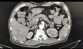
Figure 1: Aorta CT scan.
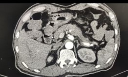
Figure 2: Celiac trunk artery.
The patient had a enteroscopy to determine the cause of their symptoms. During the procedure, it was found that there was erosion and bleeding in the small intestine due to a vascular lesion. After receiving symptomatic treatment and showing improvement, the patient was discharged. However, soon after, the patient returned to the hospital with blood in their stool. A repeat CT scan of the entire abdomen was performed, and the results were consistent with the previous findings, showing no significant changes.
Following symptomatic treatment, the patient was discharged with relief from symptoms and referred to another tertiary care facility for additional care. Due to bleeding, laparoscopic exploration and intraoperative enteroscopy were conducted. During the procedure, it was observed that the mucous membrane of the ascending colon near the hepatic flexure was swollen, blistered, and bleeding, with no clear pulsating source of the bleeding. After using endoscopic methylene blue for localization and further examination, it was determined that the bleeding might originate from a lesion in the ascending colon near the hepatic flexure, leading to the decision to perform a “right hemicolectomy and colostomy.”
After surgery, the patient experienced ongoing episodes of bloody stools from the fistula, with the bleeding attributed to intestinal vascular malformations. He was transferred to another hospital, where an abdominal trunk arteriogram (Figure 3) revealed no significant bleeding sources. Despite continued treatment for intestine bleeding bleeding related to the vascular malformation, the patient’s symptoms did not improve significantly. Consequently, a second abdominal trunk arteriogram as conducted, during which fluctuations in the patient’s blood pressure led to the identification of bleeding points. Intraoperatively, splenic artery embolization was performed, and the patient was ultimately diagnosed with a pseudoaneurysm of the splenic artery.
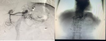
Figure 3: Abdominal trunk arteriogram.
After the surgery, the patient did not have any blood in their stool, and routine examinations did not reveal any notable issues.
Discussion
The splenic artery aneurysm ranks as the third most prevalent intra-abdominal aneurysm, following abdominal aortic and iliac artery aneurysms [1]. It is considered relatively uncommon in clinical settings, and its development is thought to be associated with abnormalities in the arterial wall structure and changes in endocrine levels [2]. Most unruptured splenic artery aneurysms do not present noticeable symptoms. Research indicates that the mortality rate for ruptured splenic artery aneurysms can be as high as 75% to 80% [3]. These aneurysms can be classified into true aneurysms and pseudoaneurysms based on the condition of the vessel wall. True aneurysms have an intact wall, whereas pseudoaneurysms occur when there is a tear in the vessel wall, allowing blood to enter a pseudo lumen and leading to the formation of a hematoma around the artery, thus compromising the vessel wall’s integrity [4]. The rupture risk for true aneurysms is estimated at 2% to 3%, while pseudoaneurysms have a rupture rate that can reach 37% to 47%, with an untreated mortality rate of 90% [5].
Rupture and bleeding from a splenic artery aneurysm can occur in various areas such as the pancreatic duct, abdominal cavity, retroperitoneum, stomach, or transverse colon. Symptoms differ based on the location of the ruptured blood vessels, with common signs including abdominal pain (29.5%), bloody or black stools (26.2%), bleeding from the pancreatic duct (20.3%), and vomiting blood (14.8%) [6]. These symptoms are often mistaken for gastrointestinal bleeding, and cases of intestine bleeding bleeding due to splenic artery aneurysm rupture are quite rare. When such a rupture occurs, it typically presents as intermittent bleeding, which can be easily overlooked or misdiagnosed. The nonspecific nature of the symptoms makes diagnosis challenging. Diagnostic methods for splenic artery aneurysms include abdominal plain films, color Doppler ultrasound, MRA, and arteriography, with CTA being the preferred option for hemodynamically stable patients with ruptured aneurysms due to its convenience, clarity, and noninvasive approach [7].
Arteriography is considered the most reliable method for diagnosing visceral aneurysms, as it provides detailed information about the aneurysm’s precise location, size, and surrounding structures, while also allowing for immediate interventional treatment. With improvements in interventional diagnostic and therapeutic techniques, percutaneous vascular intervention has become increasingly popular in clinical settings due to its benefits, including reduced trauma, shorter procedure duration, ease of use, faster recovery, and fewer postoperative complications. It is often the preferred option for treating splenic artery aneurysms [8].
Conclusion
In clinical practice, when patients experience unexplained recurrent small intestine bleeding, it is important to consider the potential for bleeding from a visceral aneurysm after ruling out more common causes like intestinal polyps, diverticula, inflammatory bowel disease, tumors, and ischemic bowel disease. Timely diagnosis and treatment of ruptured splenic artery aneurysms are vital for patient survival, and as volunteer clinicians, we must identify these cases early and take action as soon as possible.
References
- Miu YD, Ye B. Intragastric rupture of splenic artery aneurysms: three case reports and literature review. Pakistan Journal of Medical Sciences. 2013; 29: 1–4.
- Long JY, Xu D, Xie XP. Clinical diagnosis and treatment of splenic artery aneurysm: a report of 27 cases. Journal of Hepatopancreatobiliary Surgery. 2021.
- Pasha SF, Gloviczki P, Stanson AW, et al. Splanchnic artery aneurysms. Mayo Clinic Proceedings. 2007; 82: 472–9.
- Akbulut S, Otan E. Management of giant splenic artery aneurysm: comprehensive literature review. Medicine (Baltimore). 2015; 94: e1016.
- De Silva WSL, Gamlaksha DS, Jayasekara DP, et al. A splenic artery aneurysm presenting with multiple episodes of upper gastrointestinal bleeding: a case report. Journal of Medical Case Reports. 2017; 11: 123.
- Tessier DJ, Stone WM, Fowl RJ, et al. Clinical features and management of splenic artery pseudoaneurysm: case series and cumulative review of literature. Journal of Vascular Surgery. 2003; 38: 969–74.
- Al LT, Yf LG. A case of spontaneous splenic artery aneurysm rupture. Clinical Hepatology. 2019; 35.
- Panzera F, Inchingolo R, Rizzi M, et al. Giant splenic artery aneurysm presenting with massive upper gastrointestinal bleeding: a case report and review of literature. World Journal of Gastroenterology. 2020; 26: 3110–7.